
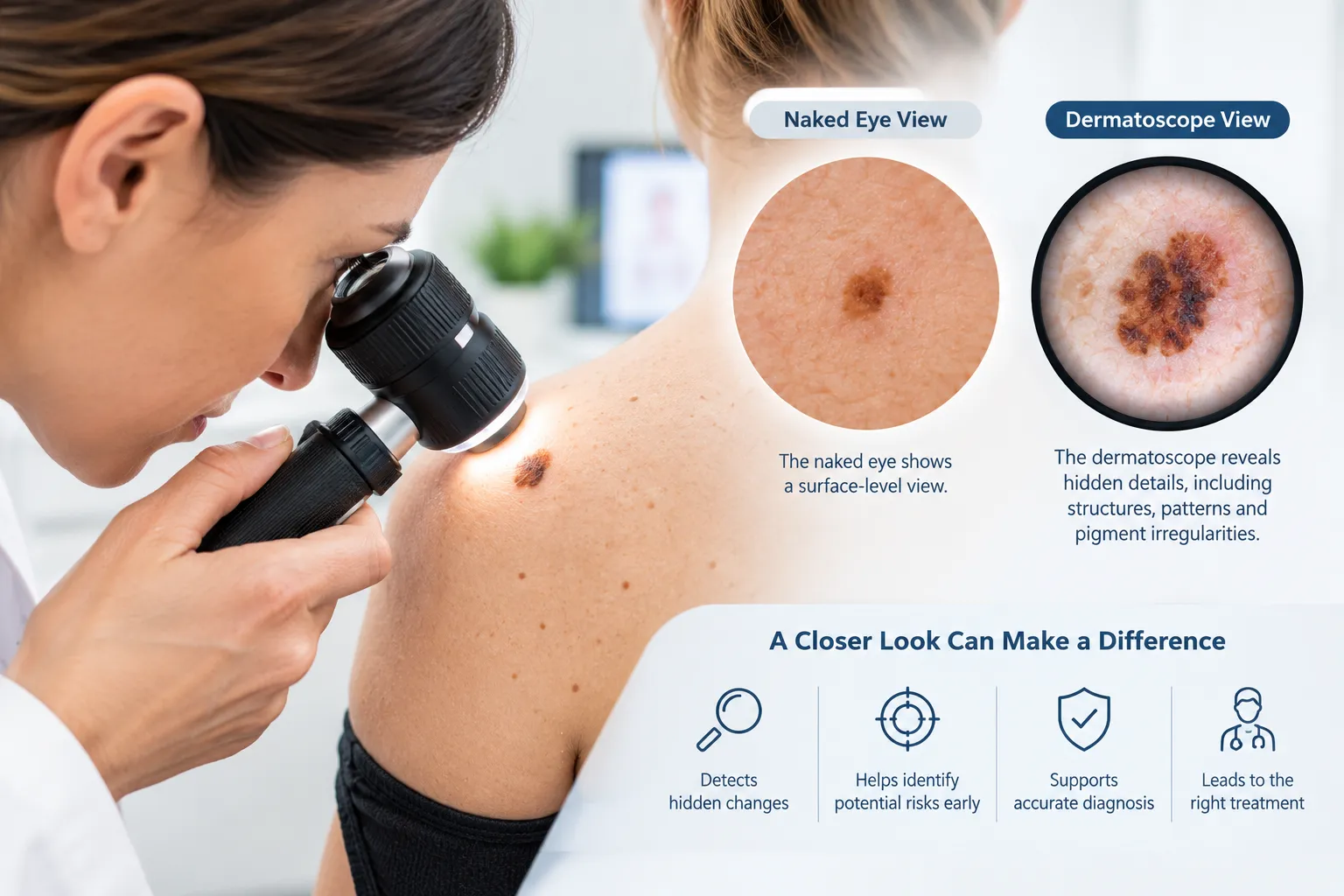
A skin check often looks simple from the patient’s side of the room. The doctor picks up a small handheld device, rests it against your skin and studies a spot you may never have given a second thought. That device is a dermatoscope, and it gives the doctor a view of your skin that careful looking alone could never provide.
Key Takeaways
- A dermatoscope reveals details beneath the skin’s surface that are difficult or impossible to see with the naked eye, including pigment patterns and blood vessel structures.
- Dermatoscopy can help distinguish suspicious lesions, allowing doctors to assess features that may indicate melanoma or other skin cancers.
- It improves the accuracy of skin examinations, particularly when assessing unusual, changing or difficult-to-classify moles and spots.
- Dermatoscopes can support earlier detection, helping identify subtle changes that may otherwise be overlooked during a standard visual examination.
- Professional assessment remains important, as dermatoscopy is a diagnostic aid that should be interpreted by an appropriately trained medical professional.
Where the Naked Eye Falls Short on Melanoma
A close look from a trained doctor might seem like enough to tell whether a spot is safe. In practice, visual assessment has firm limits, particularly with melanoma.
Cancer Council Australia reports that naked-eye clinical diagnosis of melanoma is accurate only around 60% of the time. That leaves plenty of room for error in both directions. Some cancers looking ordinary enough might slip through, and some harmless spots looking unusual enough might raise concern.
None of this reflects poorly on the doctor. It comes down to what the surface of the skin can show. Colour, outline and size offer useful clues, but much of the detail that separates a harmless mole from a cancerous one sits just beneath the surface, out of reach of the unaided eye.
How a Dermatoscope Uncovers Hidden Patterns
A dermatoscope pairs magnification with a light source that cuts down surface glare. With the glare removed, a clinician can look into a mole or lesion and see structures that stay invisible without the device. These include the way pigment spreads through the spot, the form and layout of tiny blood vessels, and fine variations in colour that help show whether a spot is behaving normally.
Skin cancers, particularly melanoma, often develop irregular internal patterns before anything looks obviously wrong on the surface. A spot that seems unremarkable can show a clearly disordered structure once it is magnified and properly lit.
The numbers show how big the gain is. According to the same Cancer Council Australia data, dermatoscopy has an accuracy of 89% and a sensitivity of 82.6% for detecting melanocytic lesions, which are spots that develop from the skin’s pigment cells. Melanoma belongs to this group, so the jump from 60% with the naked eye to 89% with a dermatoscope shows how much more a magnified, well-lit view can pick up. Dermoscopy is now a standard part of a thorough skin examination, and figures like these show why.
Why Spotting a Change Isn’t the Same as a Diagnosis
Regular self-checks are a worthwhile habit, and the evidence backs that up. A Queensland study cited by Cancer Council Australia found that nearly half (44%) of people with a confirmed melanoma diagnosis had detected the spot themselves. That figure is a strong reason to stay familiar with your own skin and to take notice when something changes.
Still, noticing a change and knowing what it means are two different things. You can see that a spot has changed, but you have no way of seeing what is going on beneath it, and that hidden layer is exactly where a dermatoscope does its work. Your own awareness and a professional examination work best as a pair, with you catching the change and the melanoma specialist confirming what it means.
How Dermoscopy Determines Your Skin Check Schedule
A dermatoscope reveals details that self-monitoring cannot reach, so professional checks are best seen as a partner to your at-home skin checks. They are especially useful for:
- spots in places you cannot easily see, such as your back
- moles you have had for years that may have changed slowly
- new spots that do not look alarming but have never been properly assessed
How often you book in depends on your personal risk factors, including your skin type, family history and past sun exposure. Your doctor can recommend a schedule that suits your level of risk.
Dermoscopy can also spare you unnecessary worry. Plenty of ordinary moles look a little irregular to an untrained eye. When a doctor can see the structure underneath, they can often confirm that a spot is benign without sending it off for a biopsy, which brings peace of mind for patient and doctor alike.
Get Every Spot Seen Under the Dermatoscope
If your last skin examination was little more than a quick glance, now is a good time to book a more thorough one. A skin cancer clinic that uses dermoscopy as standard can give you a far clearer picture of your skin than any self-check at home. A skin cancer check carried out with a dermatoscope means every spot that deserves a closer look gets one, using a tool designed to see what the naked eye cannot.
Frequently Asked Questions
Does a dermatoscope hurt or require any special preparation?
No, a dermatoscope does not hurt and needs no preparation. The doctor holds the small device gently against your skin, sometimes after applying a little gel or liquid, and the whole process is painless.
Can a dermatoscope replace the need for a biopsy?
Not completely. A dermatoscope greatly improves diagnostic accuracy and can help a doctor confirm that a spot is harmless. If a lesion still looks suspicious after examination, a biopsy is the only way to reach a definitive diagnosis.
Why did my naked-eye check miss something a dermatoscope found?
A naked-eye check is accurate for melanoma only around 60% of the time, while dermatoscopy reaches 89% accuracy for detecting melanocytic lesions, the group of pigment-cell spots that includes melanoma. Many warning patterns sit beneath the visible surface of the skin, and only magnification combined with specialised lighting can bring them into view.
Should I still check my own skin if I get regular professional checks?
Yes, keep checking. Appointments happen at set intervals, but you can look over your skin any time, so you are well placed to pick up a spot that is new or starting to change between visits. In a Queensland study, nearly half (44%) of people with confirmed melanoma were the ones who found it first.
How often should I get a dermatoscope-based skin check?
The right frequency depends on your personal risk factors, such as skin type, family history and past sun exposure. Many people benefit from an annual check, but your doctor can recommend a schedule tailored to you.
