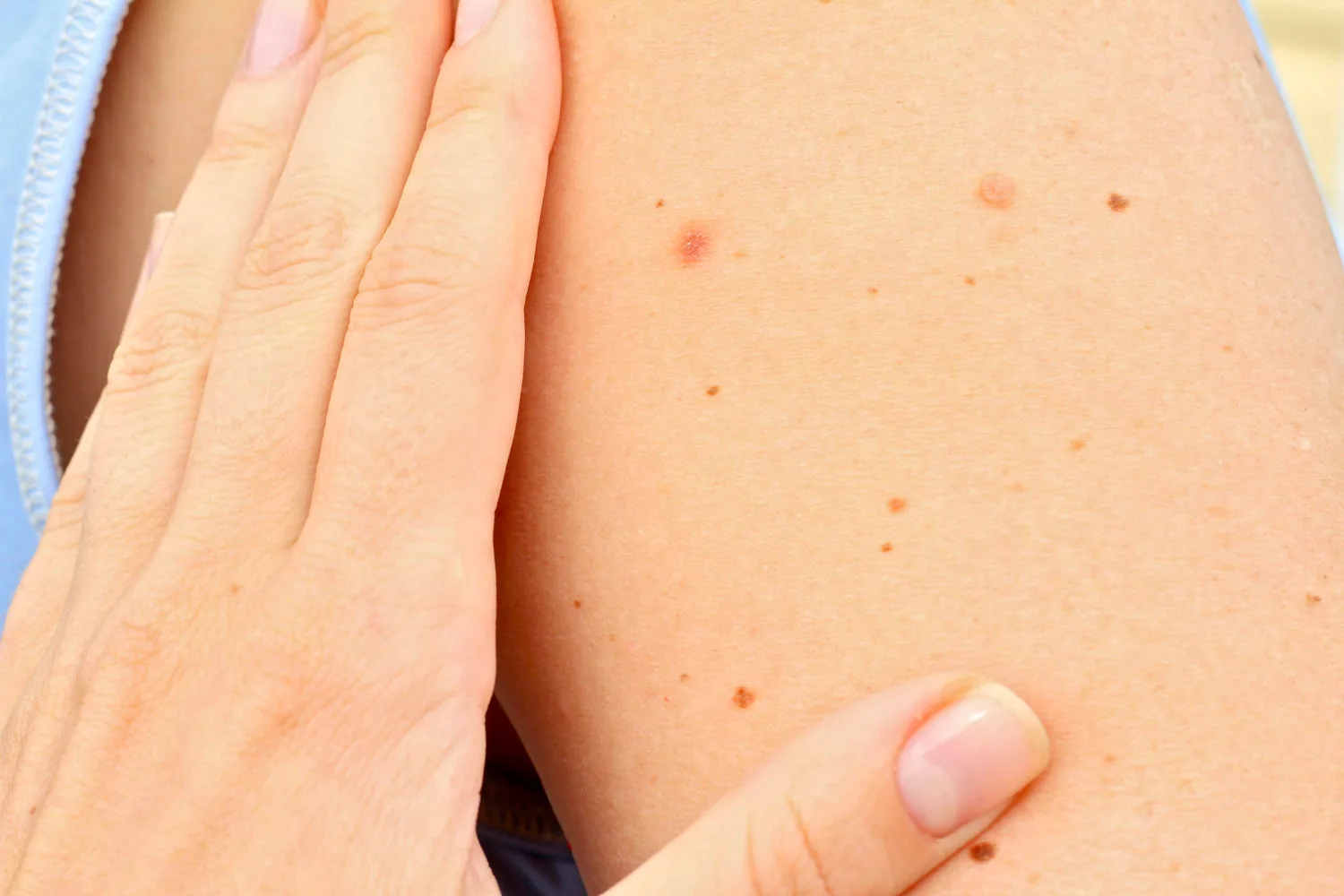
A mole can sit quietly on your skin for years, then start changing in a way that is easy to miss. That is what makes skin checks so important. Some changes are obvious, such as a mole that grows, darkens, bleeds, or looks different from the others. Other changes are more subtle, especially if they happen slowly or appear in areas you rarely check yourself.
Instead of relying only on memory or a quick look at one appointment, mole mapping creates a detailed visual record of your skin. It gives doctors a clearer way to monitor those changes over time.
Key Takeaways
- Mole mapping creates a detailed photographic baseline of skin to track moles and detect changes accurately over time.
- Early melanoma detection improves survival; Cancer Australia and National Cancer Control Indicators (NCCI) show stage 1 survival around 99%.
- Uses total body photography and dermoscopy imaging to record moles, compare changes, and detect subtle differences over time.
- Best for people with many moles, atypical lesions, fair skin, sun exposure history, or previous skin cancer risk.
- Frequency varies by risk; RACGP recommends six-monthly checks for high-risk patients, supported by Cancer Council Australia guidance.
Why Early Melanoma Detection Matters
Melanoma is one of the more serious forms of skin cancer because it can spread to other parts of the body if it is not found and treated early. Cancer Australia reports that 17,443 new cases of melanoma were diagnosed in 2025, and a person has a 1 in 19 risk of being diagnosed with melanoma of the skin by the age of 85.
When melanoma is found early, outcomes are much better. According to National Cancer Control Indicators (NCCI), stage 1 melanoma has at least 99% relative survival at five years from diagnosis.
That is why regular skin checks are not only for spots that look alarming. They are also for checking the spots you cannot see properly, including your back, scalp, soles of the feet, between the toes, and under the nails.
How Early Checks Can Improve Treatment Options
When melanoma is detected early, treatment is often more straightforward. A suspicious spot may be removed and tested before it has had the chance to spread. When detection happens later, treatment can become more complex and may involve wider surgery or additional therapies.
NCCI data show very high five-year relative survival for stage 1 melanoma, while survival decreases as melanoma becomes more advanced. That does not mean every changing mole is dangerous, but it does mean changes should not be ignored.
If you notice a new spot, a mole that looks different from your other moles, or a change in colour, shape, size, border, or texture, it is worth having it checked. A melanoma specialist can assess your skin properly and may use technology, including mole mapping and dermoscopy, to decide whether it should be monitored, reviewed again, or removed for testing.
What Is Mole Mapping?
Mole mapping is a skin monitoring process that uses detailed imaging to record the appearance and location of moles, freckles, and other skin spots. The purpose is simple: create a baseline of your skin so changes can be spotted more accurately later. However, mole mapping does not replace a doctor’s judgement.
It gives your doctor a visual history to compare against, especially if you have many moles or a higher risk of skin cancer. It is often used alongside a full skin check, dermoscopy, and clinical assessment by a trained doctor.
How Mole Mapping Records and Tracks Your Skin
Memory is not a reliable way to track moles. Most people cannot remember exactly how a mole looked six months or a year ago, especially if they have many spots across their skin.
A visual record solves that problem. It gives your doctor a reference point and helps reduce guesswork. It can also help avoid unnecessary removals when a mole looks stable across several visits.
During a mole mapping appointment, images are taken of your skin using specialised cameras. Depending on the service, this may include full-body photography, close-up dermoscopic images, or both.
Full-body images help record the overall pattern of spots across your skin. Close-up images allow doctors to examine selected moles or lesions in greater detail. These images are stored securely and used as a comparison point at future checks.
At your next appointment, your doctor can review new images beside the earlier ones. This helps them see whether a mole has changed, whether a new spot has appeared, or whether a suspicious area needs further investigation.
What Happens During Total Body Photography?
Total body photography involves taking a series of images that show the skin across the body. These images are usually captured from different angles so the doctor has a more complete record of your skin.
You may be asked to change into appropriate clothing or underwear so the images can cover the areas that need to be monitored. Although this can feel uncomfortable for some people, the purpose is medical. Cancer Council Australia recommends checking the whole body because skin cancers can appear in areas that receive little or no sun exposure.
The images are then stored and used for comparison at future visits. If a new spot appears or an existing spot changes, your doctor can compare it against your earlier photographs and decide whether further assessment is needed.
What Types of Mole Mapping Are Available?
Not every mole mapping service is the same. Some focus on total body photography, while others use dermoscopic imaging for selected moles that need closer monitoring.
At our skin cancer clinic, the most useful approach depends on your skin and your risk level. Someone with one or two spots of concern may only need targeted imaging, while someone with many moles, atypical moles, or a history of melanoma may benefit from a more complete record of their skin.
The main point is not how many photos are taken. It is whether the images help your doctor monitor your skin in a structured and clinically useful way.
How Mole Mapping Technology Finds Small Skin Changes
Mole mapping technology is designed to make skin monitoring more accurate and consistent. High-resolution photography can capture details that may be difficult to describe in notes alone, whereas dermoscopic imaging can show structures beneath the skin surface that are not visible to the naked eye.
The process usually involves three parts:
- Capturing clear images of the skin
- Reviewing selected moles or lesions in greater detail
- Comparing current images with earlier records to identify changes
This becomes especially helpful when changes are small. A mole may not look dramatically different from one visit to the next, but side-by-side image comparison can help reveal subtle shifts in size, colour, border, or pattern. Mole mapping gives your doctor more context than a single skin check can provide.
At follow-up appointments, new images can be compared with earlier ones. Your doctor may look for new lesions, changes in existing moles, or patterns that suggest a spot needs closer attention.
How AI Can Support Skin Image Review
Some mole mapping systems use artificial intelligence to assist with image review. AI can help analyse skin images, flag changes, and support comparison between current and previous records.
This does not mean AI makes the diagnosis on its own. A doctor still needs to assess the skin, consider your risk factors, review the images, and decide what should happen next.
AI is an additional tool that can help organise and analyse visual information. Used alone, without clinical judgement, it is not enough. Used well, it can support earlier recognition of suspicious changes.
Who May Benefit From Mole Mapping?
Mole mapping can be helpful for many people, but it is especially useful for those with a higher risk of melanoma. This may include people with many moles, unusual-looking moles, a personal history of skin cancer, or a family history of melanoma.
Other risk factors can include fair skin, light hair, a tendency to burn, high sun exposure, and a history of severe sunburn. People who find it difficult to monitor their own skin may also benefit, particularly if they have spots on the back, scalp, or other areas that are hard to see.
Mole mapping can also be useful if you feel unsure about whether your skin is changing. A proper baseline can make future checks clearer and less reliant on memory.
How Often Should You Have Mole Mapping?
The right timing depends on your risk level and your doctor’s advice. Some people may only need occasional imaging, while others need regular follow-up.
The Royal Australian College of General Practitioners (RACGP) recommends that people at very high risk of developing a new primary melanoma should have regular clinician checks, with six-monthly full skin examination supported by total body photography and dermoscopy. For people with lower risk, your doctor may recommend a different schedule.
Mole mapping gives your doctor a detailed record, but you are still the person most likely to notice a new spot in everyday life. If something changes before your next appointment, do not wait for the scheduled check. Have it assessed.
Take the Next Step With Your Skin Checks
Mole mapping gives your doctor a clearer way to monitor your skin over time. It creates a visual record, supports early detection, and helps track changes that may not be obvious in a single appointment. For people with many moles, previous skin cancer, unusual moles, or other melanoma risk factors, it can be a valuable part of a regular skin check routine.
If you have noticed a changing mole, a new spot, or a mark that looks different from the others, contact a reputable skin clinic and ask whether mole mapping is suitable for you.